Split-Thickness Skin Graft
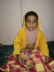
An adorable five-year-old boy and his lovely mother entered the clinic on the very first day of the camp. The boy had been badly burned around his neck and back after his shirt had caught on fire. He had second-degree burns on his back and third-degree burns around his neck and shoulders.
It was clear to the surgical team that without a skin graft that the skin would not heal easily or in a uniformed manner. Dr. Joe Bardenheier quickly stepped up to the task of completing a Split-Thickness Skin Graft.
Skin transplanted from one location to another on the same individual is termed an autograft. These grafts consist of the entire epidermis and a dermal component of variable thickness. If the entire thickness of the dermis is included, the appropriate term is Full-Thickness Skin Graft. If less than the entire thickness of the dermis is included, this graft is referred to as a Split-Thickness Skin Graft. In this case, with a large burn area and such a small boy a Split-Thickness Skin Graft was the only alternative.
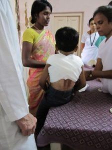
The boy and his mother entered the camp on day one to be examined.
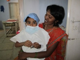
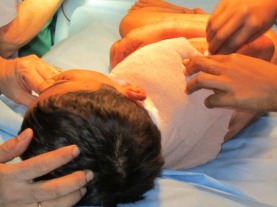
The next day the brave boy and his mother wait patiently in the pre-op area getting ready for his procedure.
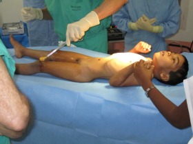
Split-thickness skin grafts may be harvested from any surface of the body, but the sites chosen should be concealed easily in recreational clothing and minimize the discomfort during reepithelialization. Common sites include the upper anterior and lateral thighs. In this case, Dr. Joe chose to take the graft from the boys left thigh.
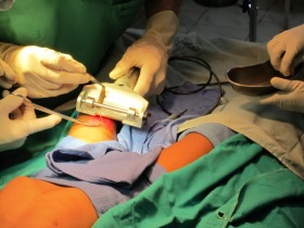
Split-thickness skin grafts may be harvested in a variety of ways. The most commonly used technique involves a dermatome, which provides rapid, consistent harvest of large uniform-thickness grafts. Dermatomes are typically air-powered or electric, although manually operated devices exist. Dermatomes harvest skin with a rapidly oscillating blade. The thickness is easily adjusted on the instrument. The width is typically adjusted in 1- to 2-inch increments by applying blade guards of different widths. The length is dictated by the surgical technique. The dermatome is a rather expensive machine and we are very thankful for its donation to the Free Surgical Clinic last year.
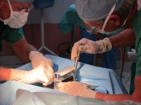
Once harvested, a split skin graft may be meshed by placing the graft on a carrier and passing it through a meshing device. The carrier has a grooved side that must be directed superiorly and upon which the graft should be laid out. This technique allows expansion of the graft surface area up to 9 times the donor site surface area. This technique is indicated when insufficient donor skin is available for large wounds, as in major burns or when the recipient site is irregularly contoured and adherence is a concern. In this case, Dr. Joe and Dr. Bob expanded the split graft to 3 times its size.
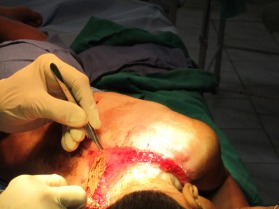
Once the recipient site has been prepared, the graft may be placed over the wound bed. Placing the dermal, typically white or lighter color, side down is important.
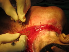
Taking care to prevent wrinkling or excessive stretching of the graft is important. Expansion slits allow wound fluid to escape through the graft rather than accumulating beneath the graft and preventing adherence. Expansion slits must heal by re-epithelialization and may contract significantly.
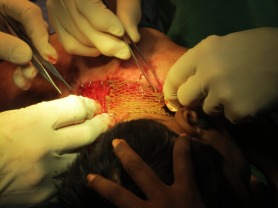
The graft must then be secured in place to provide stability during initial adherence and healing. This is most often accomplished by suturing or stapling the graft to the surrounding wound bed.
A dressing is then chosen to provide uniform pressure over the entire grafted area through a nonadherent, semiocclusive, absorbent dressing material. These dressings are meant to immobilize the graft, prevent shearing, and prevent seroma or hematoma formation beneath the graft. “Tie-over” bolster dressings are useful over joints or other areas where motion is difficult to avoid.
After graft placement, an initial adherence to the wound bed via a thin fibrin network temporarily anchors the graft until definitive circulation and connective-tissue connections are established. This adherence begins immediately and is probably maximized by 8 hours postgrafting. The period of time between grafting and revascularization of the graft is referred to as the phase of plasmatic imbibition. The graft imbibes wound exudate by capillary action through the spongelike structure of the graft dermis and through the dermal blood vessels. This prevents graft desiccation, maintains graft vessel patency and provides nourishment for the graft. This process is entirely responsible for graft survival for 2-3 days until circulation is re-established.

This young boy was truly an inspiration to everyone who came in contact with him. He exuded absolute bravery, love and kindness through-out this very painful process. We are so proud of him and very happy to have had the chance to help him.